
Cross section animal cell structure detailed Vector Image
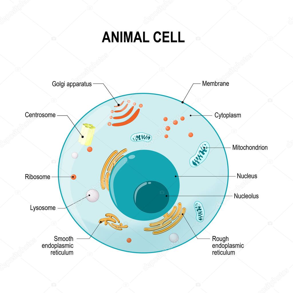
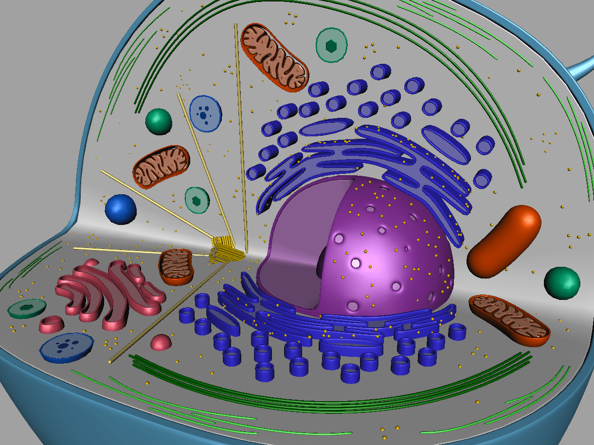
Animal cells contain a wide variety of parts, each of which plays a vital role in the survival of the cell. The Nucleus. The nucleus is the control center of the cell and houses all of the cell's genetic information. Usually, a cell has a single nucleus that contains all of its DNA molecules, but some (such as skeletal muscle cells) have more.
PPT Plant and Animal cells PowerPoint Presentation, free download ID7060098
Animal cells are eukaryotic cells, meaning they possess a nucleus and other membrane-bound organelles. Unlike plant cells, animal cells do not have cell walls, allowing for more flexibility in shape and movement. A plasma membrane encloses the cell contents of both plant and animal cells, but it is the outer coating of an animal cell.
Animal Cell Cross Section Science 3D Models Biological Science Picture Directory
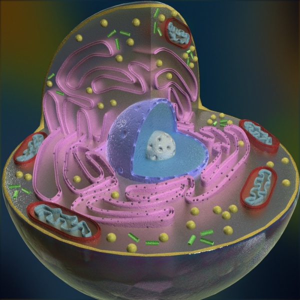
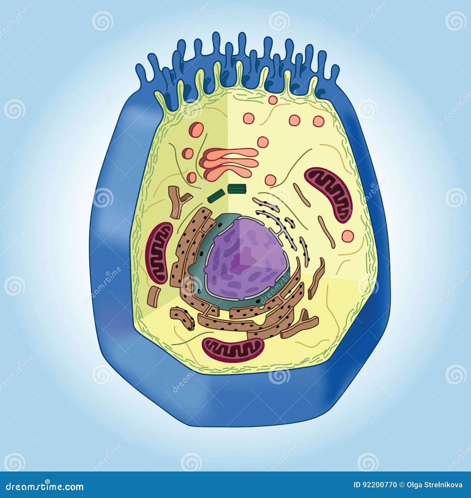
Animal Cell Diagram and Structure (cross section of an animal cell) The cell of the animal is composed of a variety of structural organelles that are enclosed within the plasma membrane. These organelles allow it to function effectively by triggering mechanisms that are beneficial to those who are the hosts (animal). The interaction of all.
Crosssection of Animal Cell on Blue Background, Structure Stock Vector Illustration of
Animal cell size and shape. Animal cells come in all kinds of shapes and sizes, with their size ranging from a few millimeters to micrometers. The largest animal cell is the ostrich egg which has a 5-inch diameter, weighing about 1.2-1.4 kg and the smallest animal cells are neurons of about 100 microns in diameter.

Animal Cell structure cross section 3D asset CGTrader
Animal cells range in size from a few microscopic microns to a few millimetres. The largest known animal cell is the ostrich egg, which can stretch over 5.1 inches across and weighs about 1.4 kilograms. This is in stark contrast to the neuron in the human body, which is just 100 microns across.
Thus, Understanding Membrane Transport, Requires An Cross Section Diagram Of An Animal Cell
For instance, in animal cell division, a ring made of actin and myosin pinches the cell apart to generate two new daughter cells. Actin and myosin are also plentiful in muscle cells, where they form organized structures of overlapping filaments called sarcomeres.. Upper: Transmission electron micrograph of flagella in cross-section, showing.
Create a cross section of an animal cell using craft supplies
Cross-Section of an animal cell. Cell Membrane. Click the card to flip 👆. A double layer that supports and protects the cell. Allows materials in and out. Click the card to flip 👆. 1 / 12.

Montessori Materials Cross Section Animal Cell Model
A site where proteins are made. Location. Term. Golgi Body (Apparatus) Definition. Modifies, processes, and packages proteins, lipids and carbohydrates. Location. Start studying Cross-Section of an Animal Cell. Learn vocabulary, terms, and more with flashcards, games, and other study tools.

Earth as a System The Eukayotic Cell
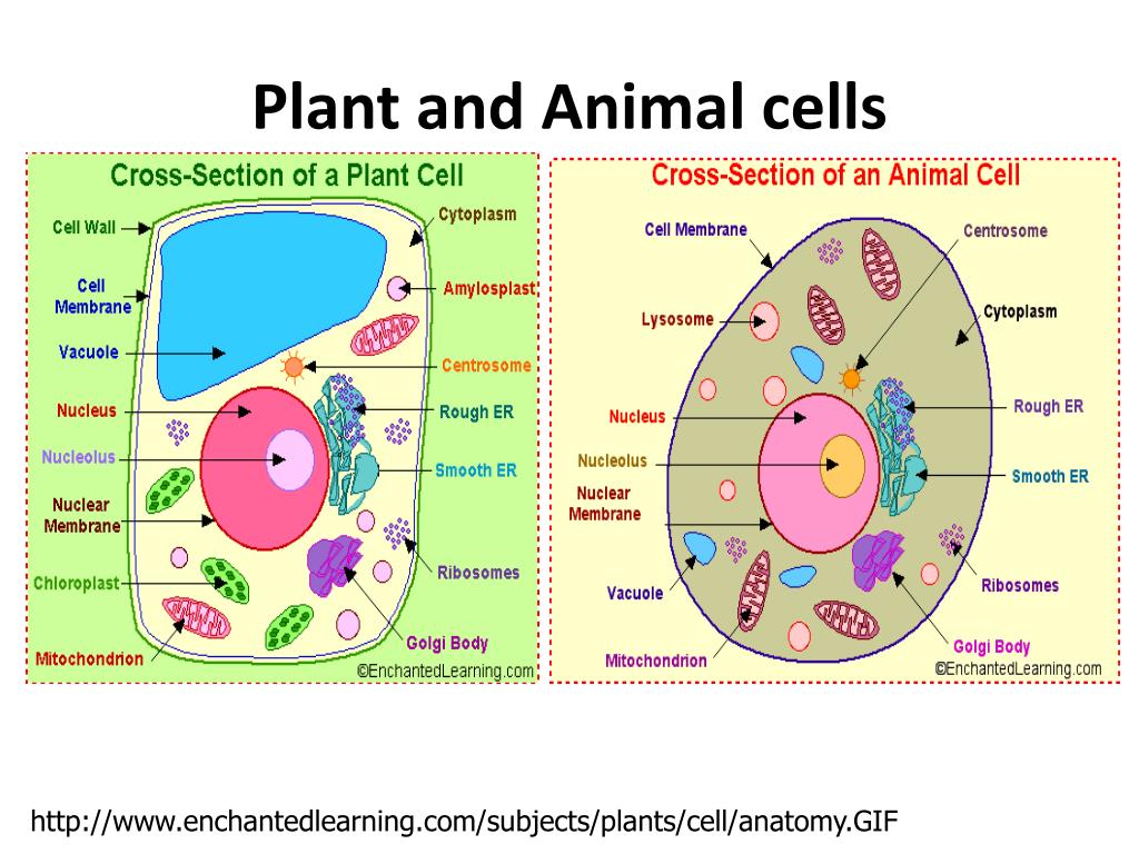
Organelles allow for various functions to occur in the cell at the same time. Despite their fundamental similarities, there are some striking differences between animal and plant cells (see Figure 1). Animal cells have centrosomes (or a pair of centrioles), and lysosomes, whereas plant cells do not. Plant cells have a cell wall, chloroplasts.

This poster is the technical illustration of cross section of Animal cell in watercolor. Each
First of all, both plants and animal cells have a cell membrane. A cell wall is more of a structural layer outside the cell membrane, mainly composed of cellulose but has other things, causing rigidity. Animals are fleshy and malleable because they lack the rigidity caused by a cell wall.
Animal Cell Structure Animal Cell Animal Cell Structure Animal Cell Images
3.1: Animal Cells. Page ID. John W. Kimball. Tufts University & Harvard. This schematic represents an idealized animal cell, e.g., a liver cell. The columns to the left and right of the labels contain links to discussions of the particular structures. Intermediate filaments.

Cross Section Animal Cell The Science Bank
A plant cell contains a large, singular vacuole that is used for storage and maintaining the shape of the cell. In contrast, animal cells have many, smaller vacuoles. Plant cells have a cell wall, as well as a cell membrane. In plants, the cell wall surrounds the cell membrane. This gives the plant cell its unique rectangular shape.

CrossSectional Animal Cell Model Carolina Biological Supply
Figure \(\PageIndex{1}\): Cross Section of a Centriole (courtesy of E. deHarven). The magnification is approximately 305,000. When a cell enters the cell cycle and passes through S phase, each centriole is duplicated. A "daughter" centriole grows out of the side of each parent ("mother") centriole.
Animal Cell 3D Model 3D Models World
This Demonstration depicts the cross section of an animal cell. Students can graphically explore the structure and relative location of each organelle as well as their unique functions.Mouseover any organelle for its name and a detailed description of its function. The control options let you select the information that shows up within the tooltip. If neither control labels are checked the toolt;

Cross Section Animal Cell Model
The cell is the basic unit of life. All organisms are made up of cells (or in some cases, a single cell). Most cells are very small; in fact, most are invisible without using a microscope. Cells are covered by a cell membrane and come in many different shapes. The contents of a cell are called the protoplasm. Glossary of Animal Cell Terms: Cell.

FileAnimal Cell Cross Section Model.jpg
Animal cells, unlike plant and fungi cells, do not have a cell wall.. As well as all the genetic material, there is also a sub-section of the nucleus called the nucleolus, which looks like a nucleus within the nucleus.. Vesicles are used extensively within the cell for metabolism and transport of large molecules that cannot cross membrane.
- Seadoo Switch For Sale Ontario
- Stainless Steel Sheets 4x8 Prices Canada
- Mr Cool Heat Pump Canada
- Once And Done By Armstrong
- Gundam Astray Red Frame Rg
- Prix Remplacement Toile Piscine Creusée
- 凡人 修仙 传 仙界 篇
- Ram 1500 Air Suspension Delete Kit
- Best Sunset Spots In Vancouver
- Ontario Security Guard Test Practice Questions